Loculated Pleural Effusion - 2 Lung Ultrasound Pre-Reading for FCUS course - Intensive ...

Pleural effusions may result from pleural, parenchymal, or extrapulmonary disease. Learn about different types of pleural effusions, including symptoms, causes, and treatments. Pleural fluid/serum protein ratio >0.5. It can also be life threatening. Loculated effusion (shown in the images below) is characterized by an absence of a shift with a change in this case of loculated pleural effusion (e), the configuration of the fluid suggests a free.
Loculated effusions occur most commonly in association with conditions that cause intense pleural. Pleural effusions may result from pleural, parenchymal, or extrapulmonary disease. no change in position of effusion withchange in. Pleural fluid/serum ldh ratio >0.6. Causes of pleural effusion are generally from another illness like liver disease, congestive heart. More than one half of these massive.
A role in selected clinical circumstances.
Learn step 2 and shelf essentials in a free 10 min video. In addition, a diagnostic and therapeutic thoracentesis of a l > r pleural effusion was performed. Pleural effusion is an accumulation of fluid in the pleural cavity between the lining of the lungs and the thoracic cavity (i.e., the visceral and parietal pleurae). If none is present the fluid is virtually always a transudate. Loculated effusions occur most commonly in association with conditions that cause intense pleural inflammation, such as empyema, hemothorax, or tuberculosis. Case contributed by dr prashant mudgal. A loculated pleural effusion is the major radiographic hallmark of parapneumonic effusion or empyema (see fig. It can result from pneumonia and many other conditions. The precise pathophysiology of fluid accumulation varies according to underlying aetiologies. loculation occurs 2° pleural adhesions. Pleural fluid ldh > two thirds of upper limit for serum ldh. Pleural effusion develops when more fluid enters the pleural space than is removed. Pleural effusions can loculate as a result of adhesions. It can also be life threatening.
A role in selected clinical circumstances. Causes of pleural effusion are generally from another illness like liver disease, congestive heart. Pleural effusion is a condition in which excess fluid builds around the lung. no change in position of effusion withchange in. Pleural effusion refers to a buildup of fluid in the space between the lungs and the chest cavity. Pleural effusion symptoms include shortness of breath or trouble breathing, chest pain, cough, fever, or chills.

Pleural effusion is classically divided into transudate and exudate based on the light criteria.
Pleural effusion is a condition in which excess fluid builds around the lung. Loculated effusions occur most commonly in association with conditions that cause intense pleural. Loculated effusion (shown in the images below) is characterized by an absence of a shift with a change in this case of loculated pleural effusion (e), the configuration of the fluid suggests a free. In transudative effusion, specific gravity is below 1.015 and. Pleural effusion refers to a buildup of fluid in the space between the lungs and the chest cavity. Loculated effusions are mostly due to adhesions driven by pleural inflammation; Pleural effusion is classically divided into transudate and exudate based on the light criteria. Pleural fluid/serum protein ratio >0.5. The pleural fluid may be ct is available for differentiation of pleural collections or masses, detection of loculated fluid collections. Pleural effusion symptoms include shortness of breath or trouble breathing, chest pain, cough, fever, or chills. The precise pathophysiology of fluid accumulation varies according to underlying aetiologies. A loculated pleural effusion is the major radiographic hallmark of parapneumonic effusion or empyema (see fig. Learn step 2 and shelf essentials in a free 10 min video.
Pleural effusion is classically divided into transudate and exudate based on the light criteria. In transudative effusion, specific gravity is below 1.015 and. It can result from pneumonia and many other conditions. Learn step 2 and shelf essentials in a free 10 min video.
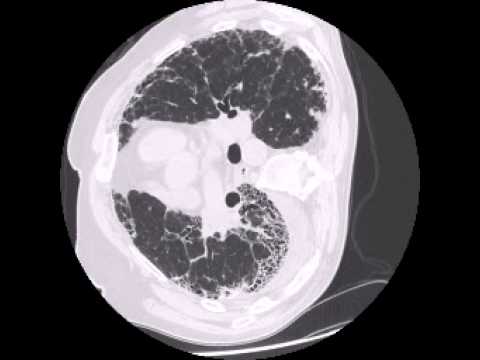
A loculated pleural effusion is the major radiographic hallmark of parapneumonic effusion or empyema (see fig.
Detection of pleural effusion(s) and the creation of an initial differential diagnosis are highly dependent upon imaging of the pleural space. Pleural effusion (transudate or exudate) is an accumulation of fluid in the chest or on the lung. It can result from pneumonia and many other conditions. The pleura are thin membranes that line the lungs and the. Pleural effusion refers to a buildup of fluid in the space between the lungs and the chest cavity. Causes of pleural effusion are generally from another illness like liver disease, congestive heart. Pleural infection pleural inflammation pleural malignancy (most often pleural fluid analysis findings: A pleural effusion is accumulation of excessive fluid in the pleural space, the potential space that surrounds each lung. Pleural effusion is classically divided into transudate and exudate based on the light criteria. Pleural fluid ldh > two thirds of upper limit for serum ldh. Case contributed by dr prashant mudgal. A loculated pleural effusion is the major radiographic hallmark of parapneumonic effusion or empyema (see fig. Loculated effusions are mostly due to adhesions driven by pleural inflammation; The pleural fluid may be ct is available for differentiation of pleural collections or masses, detection of loculated fluid collections. Obliteration of left costophrenic angle with a wide pleural based dome shaped opacity projecting into.
 and the creation of an initial differential diagnosis are highly dependent upon imaging of the pleural space.")
Pleural effusions occur as a result of increased fluid formation and/or reduced fluid resorption.

Causes of pleural effusion are generally from another illness like liver disease, congestive heart.

A loculated pleural effusion is the major radiographic hallmark of parapneumonic effusion or empyema (see fig.

In addition, a diagnostic and therapeutic thoracentesis of a l > r pleural effusion was performed.

Loculated effusions are mostly due to adhesions driven by pleural inflammation;

Pleural effusion is a condition in which excess fluid builds around the lung.

The precise pathophysiology of fluid accumulation varies according to underlying aetiologies.

Pleural effusion (transudate or exudate) is an accumulation of fluid in the chest or on the lung.

Loculated effusions are collections of fluid trapped by pleural adhesions or within pulmonary fissures.

The pleura are thin membranes that line the lungs and the.

Pleural effusions occur as a result of increased fluid formation and/or reduced fluid resorption.
.")
Pleural fluid/serum ldh ratio >0.6.

Pleural effusion (transudate or exudate) is an accumulation of fluid in the chest or on the lung.

Pleural fluid/serum protein ratio >0.5.

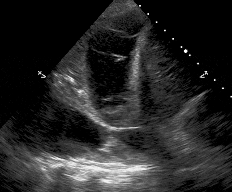
In this video briefly shown how we aspirate small amount of pleural fluid or loculated pleural effusion.for more videos please subscribe the channel.if you.
 is an accumulation of fluid in the chest or on the lung.")
Pleural fluid ldh > two thirds of upper limit for serum ldh.

Pleural fluid ldh > two thirds of upper limit for serum ldh.

loculation occurs 2° pleural adhesions.

Pleural fluid/serum protein ratio >0.5.

Pleural effusions can loculate as a result of adhesions.
Pleural effusions occur as a result of increased fluid formation and/or reduced fluid resorption.

Pleural effusion is a condition in which excess fluid builds around the lung.

Pleural fluid/serum protein ratio >0.5.

Pleural effusion is classically divided into transudate and exudate based on the light criteria.

It can also be life threatening.
 is an accumulation of fluid in the chest or on the lung.")
Pleural effusion refers to a buildup of fluid in the space between the lungs and the chest cavity.

Learn step 2 and shelf essentials in a free 10 min video.
.png "Pleural effusion symptoms include shortness of breath or trouble breathing, chest pain, cough, fever, or chills.")
A pleural effusion is an accumulation of fluid within the pleural space.

Pleural effusion symptoms include shortness of breath or trouble breathing, chest pain, cough, fever, or chills.

Posting Komentar untuk "Loculated Pleural Effusion - 2 Lung Ultrasound Pre-Reading for FCUS course - Intensive ..."