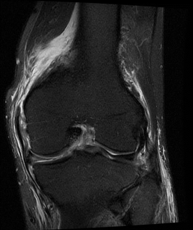
Knee Muscle Anatomy Mri / Normal Magnetic Resonance Imaging Anatomy Of The Capsular Ligamentous Supporting Structures Of The Knee Sciencedirect
Weak adductor muscles may cause knee instability and adductor strain (2). Über 80% neue produkte zum festpreis; In conclusion, we describe the normal mri anatomy of the distal biceps femoris and the relationship of this muscle with the common peroneal nerve. Mri knee anatomy scroll using the mouse wheel or the arrows. Articular muscle of the knee (articularis genu m.) normal mr imaging anatomy of the knee. When a muscle has different orientations of the tendons it means that there are different patterns of edema possible depending on the tendon injured. Anatomy basic knee mri checklist. There is a flat area of tendon originating from the knee. These motions of the knee allow the body to perform such important movements as walking, running, kicking, and jumping. Atlas of knee mri anatomy.
Superiorly, it extends to the level of the crossing of the biceps femoris tendon, and remains superficial to fcl in this location.10 There is a flat area of tendon originating from the knee. These motions of the knee allow the body to perform such important movements as walking, running, kicking, and jumping. Anatomy arthrogram anatomy basic shoulder mri. Anatomical structures of the lower limb (hip, thigh, knee, leg, ankle and foot) and specific regions (compartment of the lower. Both the pronounced accuracy of the mri and the high prevalence of knee disorders, makes the knee mri the most frequently ordered imaging procedure of the musculoskeletal system. Intensity corresponds to a pathologic lesion.

Weak adductor muscles may cause knee instability and adductor strain (2).
These motions of the knee allow the body to perform such important movements as walking, running, kicking, and jumping. Anatomy of the knee can be complicated and hard to understand. Related posts of muscle anatomy knee mri muscle anatomy get body smart. These muscles work in groups to flex, extend and stabilize the knee joint. Use the mouse scroll wheel to move the images up and down alternatively use the tiny arrows (>>) on both side of the image to move the images. Anatomy arthrogram anatomy basic shoulder mri. In this presentation mri anatomy biceps femoris muscle. Superiorly, it extends to the level of the crossing of the biceps femoris tendon, and remains superficial to fcl in this location.10 Stanford bone tumor ddx | iss/ssr msk lectures | search ocad cases | stanford virtual readouts stanford msk mri atlas has served over 1,000,000 pages to users in over 100 countries. Can also generate proton density images. Magnetic resonance imaging mri is the imaging modality of choice in the diagnosis of acute and chronic soft tissue chondral and occult skeletal injuries of the knee. Naturally, in order to assess pathologic knee imaging, it is necessary to know the appearance of a normal knee mri. The muscles of the knee include the quadriceps, hamstrings, and the muscles of the calf. Magnetic resonance imaging is particularly well suited for the medical evaluation of the musculoskeletal (msk) system including the knee, shoulder, ankle, wrist and elbow.
Knee muscle anatomy axial mri : The muscles of the knee include the quadriceps, hamstrings, and the muscles of the calf. These motions of the knee allow the body to perform such important movements as walking, running, kicking, and jumping. Anatomical structures of the lower limb (hip, thigh, knee, leg, ankle and foot) and specific regions (compartment of the lower. These muscles work in groups to flex, extend and stabilize the knee joint. The normal anatomy of the knee as seen on magnetic resonance. Louis, usa and the rijnland hospital in leiderdorp, the netherlands.
Related posts of muscle anatomy knee mri muscle anatomy get body smart.
Anatomical structures of the lower limb (hip, thigh, knee, leg, ankle and foot) and specific regions (compartment of the lower. Prescribe sagittal plane off axial images with line parallel to bony glenoid. Medical images from an mri allow medical professionals to distinguish body tissues, including the meniscus (shock absorbers in the knee), cartilage, tendons, and ligaments. Mri knee anatomy scroll using the mouse wheel or the arrows. When a muscle has different orientations of the tendons it means that there are different patterns of edema possible depending on the tendon injured. Use the mouse scroll wheel to move the images up and down alternatively use the tiny arrows (>>) on both side of the image to move the images. Über 80% neue produkte zum festpreis; Both the pronounced accuracy of the mri and the high prevalence of knee disorders, makes the knee mri the most frequently ordered imaging procedure of the musculoskeletal system. The knee joint is a modified hinge joint between the femur tibia and patella. Please email baodo at stanford.edu These are essential structures to evaluate in routine assessment of the knee on mri. Atlas of knee mri anatomy. Abnormal anatomy with normal signal, i.e.
These motions of the knee allow the body to perform such important movements as walking, running, kicking, and jumping. Injuries such as anterior cruciate ligament, meniscus and rotator cuff tears are all easily diagnosed when there is a firm understanding and knowledge of human anatomy. Knee muscle anatomy axial mri :

Articular muscle of the knee (articularis genu m.) normal mr imaging anatomy of the knee.
When a muscle has different orientations of the tendons it means that there are different patterns of edema possible depending on the tendon injured. Magnetic resonance imaging mri is the imaging modality of choice in the diagnosis of acute and chronic soft tissue chondral and occult skeletal injuries of the knee. While a detailed explanation of mri protocols and mr physics is beyond the scope of this text, fast spin echo (fse) mri is most commonly utilized for mri of the knee. This long muscle flexes the knee. There is a flat area of tendon originating from the knee. In one investigation, depicted only on the proton density weighted images. This mri knee sagittal cross sectional anatomy tool is absolutely free to use. Thigh muscles also protect neurovascular structures as they go through the proximal hip joint to the knee and lower leg (3). Doctors may recommend a knee mri if a patient experiences the following(3): The normal anatomy of the knee as seen on magnetic resonance. Louis, usa and the rijnland hospital in leiderdorp, the netherlands. The muscles of the knee include the quadriceps, hamstrings, and the muscles of the calf. Anatomy of the knee can be complicated and hard to understand.

Both the pronounced accuracy of the mri and the high prevalence of knee disorders, makes the knee mri the most frequently ordered imaging procedure of the musculoskeletal system.
 normal mr imaging anatomy of the knee.")
Cross sectional anatomy of the knee based on mri :

Stanford bone tumor ddx | iss/ssr msk lectures | search ocad cases | stanford virtual readouts stanford msk mri atlas has served over 1,000,000 pages to users in over 100 countries.
.")
Cross sectional anatomy of the knee based on mri :

Injuries such as anterior cruciate ligament, meniscus and rotator cuff tears are all easily diagnosed when there is a firm understanding and knowledge of human anatomy.

The common peroneal nerve typically courses downward within abundant fat posterior to the short head of the biceps femoris muscle and superficial to the lateral head of the gastrocnemius muscle, but.
 on both side of the image to move the images.>>) on both side of the image to move the images.")
The deepest layer consists of the popliteus muscle and its tendon passing.
.")
To realign the anterior cruciate ligament parallel with the sagittal imaging plane.
 the images obtained were exported to jpeg from dicom data stored on the pacs (picture archiving and communicating system).")
Anatomical structures of the lower limb (hip, thigh, knee, leg, ankle and foot) and specific regions (compartment of the lower.

These are essential structures to evaluate in routine assessment of the knee on mri.

While a detailed explanation of mri protocols and mr physics is beyond the scope of this text, fast spin echo (fse) mri is most commonly utilized for mri of the knee.

Anatomy of the knee can be complicated and hard to understand.

Knee muscle anatomy axial mri :

Superiorly, it extends to the level of the crossing of the biceps femoris tendon, and remains superficial to fcl in this location.10

This long muscle flexes the knee.

In this presentation mri anatomy biceps femoris muscle.
Use the mouse scroll wheel to move the images up and down alternatively use the tiny arrows (>>) on both side of the image to move the images.

Intensity corresponds to a pathologic lesion.
, cartilage, tendons, and ligaments.")
Anatomy arthrogram anatomy basic shoulder mri.
 on both side of the image to move the images.>>) on both side of the image to move the images.")
The muscles of the knee include the quadriceps, hamstrings, and the muscles of the calf.

These motions of the knee allow the body to perform such important movements as walking, running, kicking, and jumping.
 the images obtained were exported to jpeg from dicom data stored on the pacs (picture archiving and communicating system).")
Intensity corresponds to a pathologic lesion.

Stanford bone tumor ddx | iss/ssr msk lectures | search ocad cases | stanford virtual readouts stanford msk mri atlas has served over 1,000,000 pages to users in over 100 countries.

Use the mouse scroll wheel to move the images up and down alternatively use the tiny arrows (>>) on both side of the image to move the images.>>) on both side of the image to move the images.

Plantaris acts weakly to plantar flex the foot and flex the knee.

Medical images from an mri allow medical professionals to distinguish body tissues, including the meniscus (shock absorbers in the knee), cartilage, tendons, and ligaments.
 and specific regions (compartment of the lower.")
Louis, usa and the rijnland hospital in leiderdorp, the netherlands.

Plantaris can have variable size, but in most cases is difficult to demonstrate on routine mri studies.

This mri hip joint axial cross sectional anatomy tool is absolutely free to use.
While a detailed explanation of mri protocols and mr physics is beyond the scope of this text, fast spin echo (fse) mri is most commonly utilized for mri of the knee.

The common peroneal nerve typically courses downward within abundant fat posterior to the short head of the biceps femoris muscle and superficial to the lateral head of the gastrocnemius muscle, but.

Related posts of muscle anatomy knee mri muscle anatomy get body smart.

Posting Komentar untuk "Knee Muscle Anatomy Mri / Normal Magnetic Resonance Imaging Anatomy Of The Capsular Ligamentous Supporting Structures Of The Knee Sciencedirect"